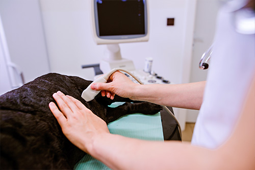
Ultrasonography (also called ultrasound or sonography) is a noninvasive, pain-free procedure that uses sound waves to examine a pet’s internal organs and other structures inside the body. It can be used to evaluate the animal’s heart, kidneys, liver, gallbladder, and bladder; to detect fluid, cysts, tumors, or abscesses; and to confirm pregnancy or monitor an ongoing pregnancy.
We may use this imaging technique in conjunction with radiography (x-rays) and other diagnostic methods to ensure a proper diagnosis. Interpretation of ultrasound images requires great skill on the part of the clinician.
We have recently purchased a new Samsung ultrasound for use in small animal abdominal imaging. This imaging technique is particularly useful for evaluating soft tissue structures that X-rays can’t clearly visualize, helping to guide treatment decisions and monitor chronic conditions.
Ultrasound does not involve radiation, has no known side effects, and doesn’t typically require pets to be sedated or anesthetized. The hair in the area to be examined usually needs to be shaved so the ultrasonographer can obtain a good result.
If you have any questions about our ultrasonography service or what to expect during your pet’s procedure, please don’t hesitate to ask.
